

Brain AVM Treatment in Guntur: Embolization and Other Options for Arteriovenous Malformation
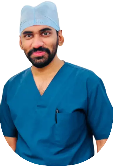
Brain AVM treatment in Guntur begins with an accurate diagnosis and a careful decision about whether treatment is needed at all. Dr. G. P. Venkat Choudary is a Consultant Interventional Radiologist at KIMS-SIKHARA Hospitals who performs catheter-based endovascular procedures, including AVM embolization, for abnormal tangles of brain blood vessels called arteriovenous malformations. Each plan is individual, and embolization is one option among several.
A sudden severe headache, a first seizure, new weakness, or trouble speaking or seeing can signal a brain bleed. Call 108 or 112 and go to the nearest hospital with emergency care and a CT scan.
- MBBS, MD Radiology, DrNB Interventional Radiology
- Consultant Interventional Radiologist, KIMS-SIKHARA Hospitals
- 5+ years of experience
What Is a Brain AVM (Arteriovenous Malformation)?
A brain arteriovenous malformation (AVM) is a tangle of abnormal blood vessels in which arteries connect directly to veins, without the fine capillaries that normally sit in between. Blood flows through this tangle, called the nidus, at high pressure, and the vessel walls can weaken and bleed. Most AVMs are thought to be present from birth, and they are uncommon. Many are found by chance on a scan done for another reason.
The risk of bleeding differs from person to person. A 2017 American Heart Association scientific statement puts the yearly risk of a first bleed from an unruptured AVM at about 1%, and the yearly risk of another bleed after a rupture at about 5%. Deep venous drainage, an aneurysm on a feeding artery and a previous bleed can raise the risk.
Brain AVM Symptoms and Who Needs Evaluation
Many brain AVMs cause no symptoms until they bleed. When symptoms appear, they may include:
- Headaches, sometimes on one side
- Seizures
- Sudden severe headache with vomiting, which may mean a bleed
- Weakness, numbness or tingling in part of the body
- Trouble with speech, vision or balance
You should be assessed if an AVM has been found on an MRI or CT scan, if you have had an unexplained seizure or a sudden severe headache, or if a known AVM causes new symptoms. A bleeding AVM is a type of haemorrhagic stroke, so sudden symptoms need emergency care. See also our page on brain stroke treatment in Guntur.
Brain AVM Embolization Treatment in Guntur: Step by Step
- Consultation: the doctor reviews symptoms, any earlier bleed, seizures, medicines and existing scans.
- Imaging: MRI and CT angiography show the AVM and the brain around it. A cerebral angiogram, a catheter test, shows the feeding arteries, the nidus, the draining veins and any aneurysm in fine detail.
- Grading and planning: the Spetzler-Martin grade scores the AVM by size, location in functionally important brain and venous drainage. The team then weighs observation, embolization, surgery, radiosurgery or a combination.
- Embolization: usually under general anaesthesia, a thin catheter is guided under X-ray from the groin or wrist to the brain. A micro-catheter is placed in the feeding arteries and a liquid embolic agent or coils block the nidus. Large or complex AVMs are often treated in several stages.
- Recovery monitoring: you are watched closely afterwards, with careful blood pressure control and neurological checks.
- Follow-up: repeat imaging shows whether more embolization, surgery or radiosurgery is needed.
Benefits and Limits of Endovascular Treatment for Brain AVM in Guntur
Endovascular treatment reaches the AVM through blood vessels, so the skull does not need to be opened. Depending on the AVM, embolization can:
- Close the AVM completely, mainly in small, low-grade lesions
- Shrink the nidus before surgery or radiosurgery, making them safer or possible
- Close a weak spot, such as an aneurysm, that is likely to bleed
- Reduce blood flow to ease symptoms in selected cases
The limits matter. A 2024 meta-analysis of 25 studies (1,425 patients) found complete closure in about half of AVMs treated with curative intent, with about a quarter needing retreatment, and concluded that curative embolization suits mainly small, low-grade AVMs. Results vary widely, and many AVMs need more than one treatment.
Brain AVM Treatment in Guntur: Options Compared
| Option | What it involves | Main advantage | Main limits |
|---|---|---|---|
| Observation and medicines | Regular imaging, medicines for headache or seizures | Avoids procedure risks | The AVM stays, so the bleeding risk remains |
| Endovascular embolization | Catheter and liquid embolic agent or coils | No skull opening, can be staged | Complete cure only in selected AVMs, may need repeat sessions |
| Microsurgery | Open surgery to remove the AVM | Can remove the AVM in one operation when it is accessible | Needs a craniotomy, risk is higher for deep or large AVMs |
| Stereotactic radiosurgery | Focused radiation to the nidus | No incision, suits small deep AVMs | Closure can take 2 to 3 years, and bleeding risk continues meanwhile |
| Combination treatment | Embolization with surgery or radiosurgery | Each step makes the next easier | Needs a coordinated team and more than one procedure |
Who May Need Treatment, and Who May Not
Treatment is not automatic. The ARUBA trial randomised people with unruptured AVMs to treatment or observation and was stopped early because death or stroke occurred in 30.7% of the treatment group versus 10.1% of the observation group at about 33 months. The gap persisted in later follow-up. Critics note that many eligible patients were not enrolled and that specialist centres report better results, so the trial is debated, but it shows why observation is reasonable for many unruptured AVMs.
Treatment is more often advised after a bleed. Other points that may favour it include an aneurysm linked to the AVM, a small low-grade AVM, worsening symptoms and seizures that are hard to control. Age, general health and personal preference also count. Where another treatment is better, you will be told so.
Risks and Safety of AVM Embolization
Embolization carries real risks. A 2025 systematic review put the overall complication rate near 10%, with bleeding complications in about 5% of cases. Possible problems include:
- Bleeding during or after the procedure
- Stroke from a blocked healthy vessel
- Seizures or new weakness, speech or vision problems
- Embolic material travelling to an unintended vessel
- Bleeding or bruising at the catheter site
- Reaction to contrast dye or risks of anaesthesia
- Remaining AVM that needs more treatment, or return of the AVM
Risks depend on the size, location and structure of the AVM, and they are discussed in detail before any procedure.
Recovery After AVM Embolization
Most people stay in hospital for observation after the procedure, and the length of stay depends on the AVM and any complications. Blood pressure control, rest and gradual return to normal activity are usual. Seizure or headache medicines may need to continue. A follow-up angiogram or MRI checks the result and decides the next step, which may be another embolization session, surgery or radiosurgery. Call emergency services at once for a sudden severe headache, a seizure, new weakness or speech trouble.
Brain AVM Treatment Cost in Guntur: What Affects the Bill
The cost of brain AVM treatment in Guntur cannot be quoted before the AVM has been imaged and graded, because plans differ widely. The main factors are:
- Imaging: MRI, CT angiography and cerebral angiogram
- Whether treatment is embolization alone or combined with surgery or radiosurgery
- The number of embolization sessions
- Catheters, balloons, coils and liquid embolic agents
- Anaesthesia, ICU and ward stay, and room category
- Complications or emergency care after a bleed
- Follow-up imaging, medicines and rehabilitation
- Insurance or government scheme coverage, which varies by hospital and policy
Ask for a written estimate that lists what is included and what could change it.
Choosing a Brain AVM Embolization Specialist in Guntur
AVM care works best as teamwork between interventional neuroradiologists or neurointerventionists, neurosurgeons and radiosurgery specialists. Useful questions to ask:
- Which treatments does the team recommend, and why not the others?
- How many brain AVMs does the team treat, and what are the risks for my AVM grade?
- Is neurosurgical and ICU support available, and can radiosurgery be arranged if needed?
- What are the chances of needing more than one procedure?
A second opinion is reasonable, and anyone who promises a guaranteed cure should be treated with caution.
Why Choose Dr. G. P. Venkat Choudary
Dr. G. P. Venkat Choudary (MBBS, MD Radiology, DrNB Interventional Radiology) is a Consultant Interventional Radiologist at KIMS-SIKHARA Hospitals, Agatha Varappadu, Guntur, with 5+ years of experience. He trained in neuro and vascular interventional radiology at Max Super Specialty Hospital, Saket, New Delhi. His listed services include AVM embolization, aneurysm coiling, intracranial stenting and stroke thrombectomy, and neurovascular interventions are among his areas of special interest. He is a life member of the Society of Therapeutic Neuro Interventions and the Indian Society of Vascular and Interventional Radiology, presented neurovascular and embolization cases at ISVIR 2023 and has received ISVIR recognition for a best paper (2022) and a scientific poster (2023). Patients can expect a plain explanation of the diagnosis, the options and the risks before any decision.
Read more about Dr. Choudary or see his neurovascular services.
Frequently Asked Questions About Brain AVM Treatment
What is the treatment for a brain AVM?
Options include observation with regular imaging, endovascular embolization, microsurgery and stereotactic radiosurgery, often in combination. The choice depends on whether the AVM has bled, its size and location, your age and symptoms, and the risks of each option.
Where can I get brain AVM treatment in Guntur?
Dr. G. P. Venkat Choudary consults at KIMS-SIKHARA Hospitals, Agatha Varappadu, Guntur, and performs endovascular procedures including AVM embolization. A sudden severe headache, seizure or new weakness is an emergency, so call 108 or 112 and go to the nearest emergency department.
Can a brain AVM be cured with embolization?
Sometimes. Embolization can close small, low-grade AVMs completely, but many AVMs need more than one session or a combination with surgery or radiosurgery. Studies show wide variation in results, so a cure cannot be promised.
Does every brain AVM need treatment?
No. Some unruptured AVMs are safely monitored, because treatment itself carries risk. Treatment is more often advised after a bleed or when features suggest a higher risk. The decision is made case by case after full imaging.
How much does brain AVM treatment cost in Guntur?
The cost depends on the imaging needed, the treatment chosen, the number of sessions, devices used, hospital stay and any complications. It cannot be quoted before the AVM is assessed. Ask for a written estimate and check insurance or scheme coverage.
What is endovascular treatment for brain AVM?
It is treated through blood vessels. A thin catheter is guided from the groin or wrist to the AVM under X-ray, and a liquid embolic agent or coils are placed to block the abnormal vessels, without opening the skull.
What are the risks of AVM embolization?
Risks include bleeding, stroke from a blocked healthy vessel, seizures, new neurological problems, catheter site problems and a remaining AVM that needs more treatment. Reviews report complications in roughly one in ten procedures.
How long is recovery after AVM embolization?
Most people are observed in hospital afterwards, and the length of stay depends on the AVM and any complications. Activity is increased gradually, and follow-up imaging decides whether more treatment is needed.
Talk to Dr. G. P. Venkat Choudary About Brain AVM Treatment in Guntur
If you or a family member has been told about a brain AVM, or needs a second opinion on brain AVM treatment in Guntur, book a consultation and bring your MRI, CT and angiogram reports. For sudden severe symptoms, call 108 or 112.
Address: KIMS-SIKHARA Hospitals, Mangalagiri Road, near Best Price, Agatha Varappadu, Guntur, Andhra Pradesh 522001
Phone and WhatsApp: +91 79936 64215
Clinic hours: Monday to Saturday, 10 AM to 5 PM (not an emergency service)
Prefer not to fill a form? Call +91 79936 64215 or message on WhatsApp.
Medical disclaimer
This page is for general information and does not replace medical advice, diagnosis or treatment. A suspected brain bleed is a medical emergency, so call 108 or 112 without delay. Treatment decisions depend on each patient's scans and condition, and results vary.
References
- Derdeyn CP, et al. Management of Brain Arteriovenous Malformations: A Scientific Statement for Healthcare Professionals From the American Heart Association/American Stroke Association. Stroke. 2017;48:e200-e224. Supports: yearly bleeding risks and risk factors.
- Mohr JP, et al. Medical management with or without interventional therapy for unruptured brain arteriovenous malformations (ARUBA): a multicentre, non-blinded, randomised trial. Lancet. 2014;383:614-621. doi:10.1016/S0140-6736(13)62302-8. Supports: 30.7% vs 10.1% death or stroke at about 33 months.
- Mohr JP, et al. ARUBA final follow-up of a multicentre, non-blinded, randomised controlled trial. Lancet Neurol. July 2020. PMID 32562682. Supports: the difference persisted at about 50 months.
- Primary embolization of cerebral arteriovenous malformations with intention to cure: a systematic review of literature and meta-analysis. Neurosurgery. 2024;95(6):1232-1244. doi:10.1227/neu.0000000000003001. Supports: about half fully closed, about a quarter retreated, suited to small low-grade AVMs.
- Sato K, Matsumoto Y, Ezura M, Endo H. Transarterial embolization for the management of brain arteriovenous malformations: a systematic review. J Neuroendovasc Ther. 2025;19(1):ra.2024-0049. Supports: complication rate near 10%, bleeding complications near 5%.
- UCSF Brain AVM Registry comparison of ARUBA-eligible patients: https://pmc.ncbi.nlm.nih.gov/articles/PMC4420142 . Supports: specialist-centre results differed from ARUBA.
- UW Medicine patient education. Treating Arteriovenous Malformations: https://healthonline.washington.edu/sites/default/files/record_pdfs/Treating-Ateriovenous-Malformations_a11y.pdf . Supports: radiosurgery closure can take 2 to 3 years.