


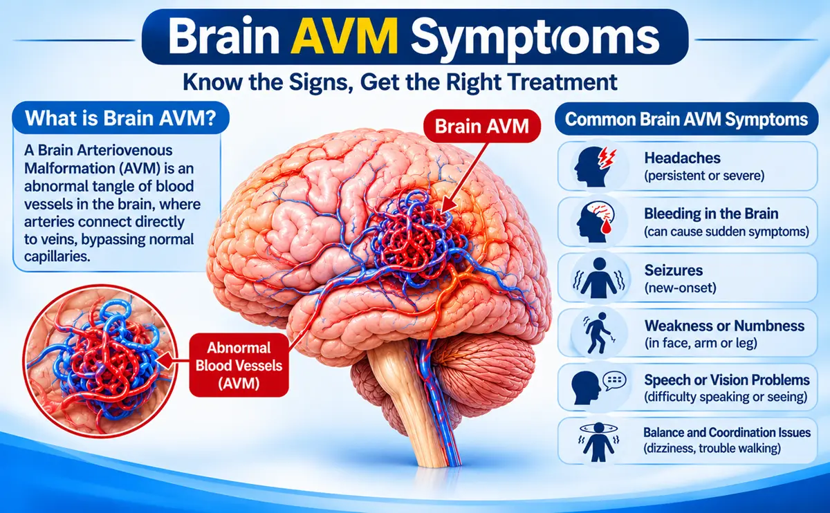
Brain AVM Symptoms and Diagnosis: When to See a Doctor
Many brain AVMs cause no symptoms for years. When they do, the usual signs are a sudden bleed (severe headache, vomiting, weakness or confusion), seizures or headaches. An AVM is suspected on an MRI or CT scan and confirmed with a cerebral angiogram. See a doctor urgently for a sudden severe headache or a first seizure, and have any AVM found by chance assessed by a specialist, because not every AVM needs treatment.
A brain arteriovenous malformation (AVM) is a tangle of abnormal blood vessels in which arteries connect directly to veins, without the fine capillaries in between. This guide covers the symptoms, the causes, how an AVM is diagnosed and when to see a doctor.
What Is a Brain AVM?
In a brain AVM, blood flows from arteries straight into veins through a knot of vessels called the nidus. The high-pressure flow can weaken the vessel walls, which may then bleed. AVMs are uncommon, with an estimated 10 to 18 cases per 100,000 people. They are usually present from birth, but symptoms most often start between the 20s and 40s. Some AVMs are discovered by chance on a scan done for another reason.
Brain AVM Symptoms
| How it shows up | How often | What it can feel like |
| Bleeding (hemorrhage) | About half of people at diagnosis | Sudden severe headache, vomiting, weakness, confusion, drowsiness |
| Seizures | Roughly 15% to 40% | Convulsions, staring spells, twitching in one arm or leg |
| Headaches | Roughly 10% to 50% | Persistent or migraine-like headaches, with no typical pattern |
| Neurological deficits | Less common | Weakness, numbness, or trouble with speech, vision or balance |
| No symptoms | A minority | Found by chance on a scan |
Figures vary between studies, so treat them as rough guides. Some symptoms come from a bleed, and others come from pressure on nearby brain or from changes in blood flow around the AVM.
Emergency Signs and Warning Signs
Call 108 or 112 at once for:
- A sudden, severe headache, often described as the worst of your life
- A first-ever seizure
- Sudden weakness, face drooping, speech trouble or vision loss
- Repeated vomiting with drowsiness, confusion or fainting
See a doctor soon for repeated seizures, headaches that change or come with other neurological symptoms, or a known AVM that causes new symptoms. A headache on its own is rarely caused by an AVM, and there is no tell-tale headache pattern. Most headaches have other causes, but a new pattern with other warning signs deserves a scan.
What Causes a Brain AVM? Is It Inherited?
The exact cause is not fully understood. Most AVMs are thought to form during development before birth, and they are generally not inherited. Research has found acquired gene changes, including KRAS mutations, in many AVMs that occur without a family history. Rarely, AVMs appear as part of inherited conditions such as hereditary hemorrhagic telangiectasia. There is no proven way to prevent a brain AVM.
Bleeding Risk: What the Numbers Mean
A 2017 American Heart Association scientific statement estimates the yearly risk of a first bleed from an unruptured AVM at about 1%, and the yearly risk of another bleed after a rupture at about 5%. Other sources quote 2% to 4% a year, and individual risk varies widely. A previous bleed, a deep location, deep venous drainage and an aneurysm linked to the AVM are factors that raise it. These are averages, not predictions for one person, which is why each AVM is assessed on its own features.
How Is a Brain AVM Diagnosed?
- Examination: the doctor asks about headaches, seizures and weakness and checks nerve function.
- CT scan: quick and very good at showing a bleed, but it may miss the AVM itself.
- MRI and MR angiography: show the nidus, the draining veins and the surrounding brain without a catheter.
- CT angiography: gives a fast picture of the vessels, which is useful in an emergency.
- Cerebral angiogram (digital subtraction angiography): a catheter test, widely regarded as the gold standard, that shows the feeding arteries, draining veins and any aneurysm in fine detail.
- Grading: the Spetzler-Martin grade scores the AVM by size, location and venous drainage to help plan care.
| Test | Main strength | Main limit |
| CT scan | Fast, shows bleeding | Can miss the AVM |
| MRI and MRA | No catheter, shows nidus and brain | Less vessel detail, small lesions can be missed |
| CT angiography | Quick vessel map | Radiation and contrast dye |
| Cerebral angiogram | Most detailed, guides treatment | Invasive, small risk of complications |
Common Myths About Brain AVMs
- Myth: every AVM will bleed. Fact: many never bleed, and the yearly risk varies widely between people.
- Myth: a headache means an AVM. Fact: AVMs are uncommon, and most headaches have other causes.
- Myth: an AVM runs in families. Fact: most are not inherited.
- Myth: AVMs only form before birth. Fact: most do, but rare reports describe new AVMs forming later in life.
- Myth: every AVM needs surgery. Fact: options include monitoring, embolization, surgery, radiosurgery or a combination, chosen case by case.
- Myth: a normal CT scan rules out an AVM. Fact: CT can miss an AVM, and MRI or angiography may be needed if suspicion remains.
What Happens After Diagnosis?
Finding an AVM does not always mean treatment. Options include regular monitoring with medicines for seizures or headaches, endovascular embolization, microsurgery, radiosurgery or a combination. The ARUBA trial found that many unruptured AVMs did better with monitoring than with intervention over the first years, although its findings are debated. A bleed, a small low-grade AVM or a linked aneurysm may tip the balance towards treatment. Our page on brain AVM treatment in Guntur compares the options.
Living With a Brain AVM
Take seizure or headache medicines as prescribed, keep follow-up scan appointments and report new symptoms quickly. Ask your doctor about pregnancy, blood-thinning medicines, strenuous exercise and driving, because advice depends on the AVM and on any seizures. A bleeding AVM is a type of hemorrhagic stroke, so it helps to know the warning signs described in our guide to ischemic vs hemorrhagic stroke.
When to See a Doctor
See a doctor if an AVM has been found on a scan, if you have had an unexplained seizure, or if you have new neurological symptoms or a changing headache pattern. Go to an emergency department straight away for a sudden severe headache, weakness, speech trouble or collapse.
Frequently Asked Questions
What are the first signs of a brain AVM?
Many people have none. When signs appear, the most common are a sudden bleed with severe headache, a seizure, headaches or weakness, numbness or speech and vision problems.
Can a brain AVM be seen on a CT scan or MRI?
Yes, often. A CT scan shows bleeding and may show a large AVM, while an MRI shows the tangle of vessels more clearly. A cerebral angiogram gives the most detail and is usually needed to plan treatment.
Is a brain AVM hereditary?
Usually not. Most brain AVMs occur without a family history. Rarely they are part of an inherited condition such as hereditary hemorrhagic telangiectasia.
What is the difference between an AVM and an aneurysm?
An AVM is a tangle of abnormal vessels linking arteries directly to veins, while an aneurysm is a weak bulge in an artery wall. Both can bleed, and an aneurysm can sometimes occur together with an AVM.
Does a brain AVM always need treatment?
No. Some unruptured AVMs are monitored because treatment carries its own risks. Treatment is more often advised after a bleed or when features suggest a higher risk. The decision is made case by case.
Is a cerebral angiogram safe?
It is an invasive test with a small risk of complications such as bleeding at the puncture site or a stroke. Doctors use it when the detail it gives is needed to plan care.
When is a brain AVM an emergency?
A sudden severe headache, a first seizure, sudden weakness, speech trouble or collapse are emergencies. Call 108 or 112 and go to the nearest hospital with a CT scan.
Where can I get brain AVM treatment in Guntur?
Dr. G. P. Venkat Choudary, an interventional radiologist, consults at KIMS-SIKHARA Hospitals, Agatha Varappadu, Guntur, and performs endovascular procedures including AVM embolization.
Talk to a Specialist
If you have been told you have a brain AVM, or want a second opinion on brain AVM treatment in Guntur, you can book a consultation with Dr. G. P. Venkat Choudary, Consultant Interventional Radiologist at KIMS-SIKHARA Hospitals, Mangalagiri Road, near Best Price, Agatha Varappadu, Guntur, Andhra Pradesh 522001. Bring your MRI, CT and angiogram reports. Call or WhatsApp +91 79936 64215 (Monday to Saturday, 10 AM to 5 PM; not an emergency service). Book an Appointment
Medical disclaimer: This article is for general information and does not replace medical advice, diagnosis or treatment. A suspected brain bleed is a medical emergency, so call 108 or 112 without delay.
References
- Derdeyn CP, et al. Management of Brain Arteriovenous Malformations: A Scientific Statement for Healthcare Professionals From the American Heart Association/American Stroke Association. Stroke. 2017;48:e200-e224. Supports: about 1% and about 5% yearly bleeding risks, risk factors.
- Mohr JP, et al. Medical management with or without interventional therapy for unruptured brain arteriovenous malformations (ARUBA): a multicentre, non-blinded, randomised trial. Lancet. 2014;383:614-621. doi:10.1016/S0140-6736(13)62302-8. Supports: monitoring did better than intervention in the first years.
- National Organization for Rare Disorders. Arteriovenous malformation: https://rarediseases.org/rare-diseases/arteriovenous-malformation/ . Supports: presentation rates, 2% to 4% yearly bleeding risk, no typical headache pattern, MRI and angiography.
- Review of brain AVM epidemiology, presentation and diagnosis, PMC4216697: https://pmc.ncbi.nlm.nih.gov/articles/PMC4216697 . Supports: prevalence of 10 to 18 per 100,000, age at first bleed, angiography as gold standard.
- ‘De Novo’ Brain AVMs: hypotheses for development and a systematic review of reported cases, PMC7996785: https://www.ncbi.nlm.nih.gov/pmc/articles/PMC7996785/ . Supports: KRAS mutations in many sporadic AVMs, rare new AVMs.
- Book chapter on intracranial AVM diagnosis, doi:10.1007/978-3-319-92306-2_22. Supports: inherited syndromes, CT and MRI findings, angiography.